
What Happens After The 2WW?
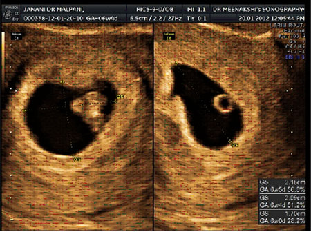
Those two weeks after fertility treatment feel like the longest days of your life. Every morning you wake up hoping for a sign, every ache or twinge makes your heart flutter with anxiety. You tell yourself not to get your hopes up, but you cannot help but wonder: What happens now? Is it working? Will this finally be the good news you have been waiting for?
The Wait is Over, But the Worry Isn’t
You finally reach the end of that dreaded two week wait (2WW), and you are told it is time to test your HCG levels. Maybe you get that first glimmer of hope: The numbers are rising. But no one really prepares you for what comes next. The truth is, after years of disappointment, you know good news does not always last—and the hardest part is that the waiting is not over yet. Now, the next phase begins: Tracking and understanding your pregnancy through ultrasounds.
Let us be honest: Ultrasound scans can be both a moment of hope and a source of dread. You stare at those blurry, black and white images, trying to make sense of the shapes, while every scan feels like another test of your emotional strength.
Making Sense of Early Pregnancy Ultrasounds: What You See, What It Means
We believe knowledge is power. At Malpani Infertility Clinic, we want you to understand what you are seeing on the ultrasound screen, so you are not left guessing or feeling powerless. Below is an atlas of real ultrasound images from early pregnancies, with explanations to help you know what is normal, what is worrisome, and what needs a follow-up.
These images are provided courtesy of N M Medical Center.
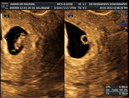
This ultrasound is from a woman 6 weeks and 4 days pregnant (almost 7 weeks). The gestational sac (GS) is visible: It is a well-defined, dark area about 2 cm in size, filled with amniotic fluid. Inside, you can see a small white ring—the yolk sac (YS)—which supports the baby in the earliest weeks. The fetus itself is about 1 cm long (measured as crown-rump length, or CRL). When you see this, it is a good sign: The pregnancy is progressing well and is on track for the dates.
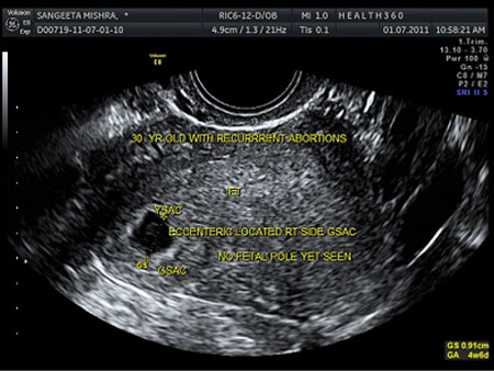
Here is an image from a 30-year-old woman with a history of miscarriages. This scan is at 5 weeks. You can spot a clear gestational sac and a yolk sac, but no fetal pole yet (the earliest sign of the embryo). At this point, it is too early to judge the health of the pregnancy. One scan is not enough, especially before 6 weeks. A follow-up scan after a week is crucial to see if the pregnancy develops as expected.
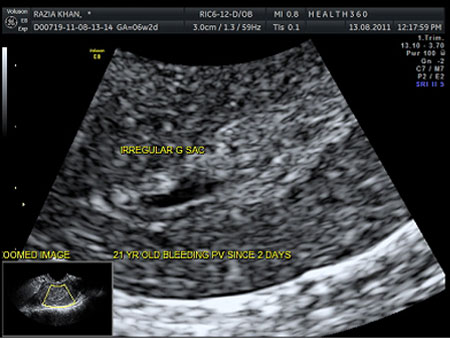
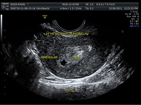
For a 21-year-old woman, 5 weeks pregnant, who had two days of vaginal bleeding, the scan tells a difficult story. The gestational sac here is irregular and collapsing. This is unfortunately a non-viable pregnancy, which will likely end as a miscarriage without medical intervention.
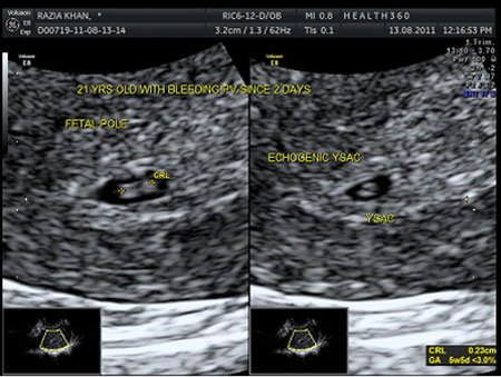
Contrast that to another 21-year-old, 6 weeks pregnant, also with two days of bleeding. This scan shows a twin pregnancy: One embryo has a visible fetal pole; the other is lagging behind with just a yolk sac. This second sac might catch up, or it could stop developing—a phenomenon called “vanishing twin.” Again, you need a repeat scan in a week to judge what is really happening.
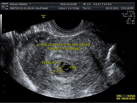
This scan belongs to a 22-year-old woman who should be 7 weeks and 6 days along, based on her dates. The gestational sac is inside the uterus and looks normal, but the fetus is much smaller than expected, measuring only 4 mm (matching just 6 weeks). In such cases, there is nothing to do but wait for another week and check again. It is a stressful waiting game, but sometimes pregnancies can surprise us and catch up.
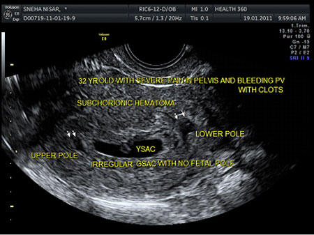
When a 32-year-old comes in at 8 weeks pregnant with severe cramps and bleeding, the scan can be truly worrying. Here, you see a subchorionic hematoma—a collection of blood in the uterus. Alone, this is not always dangerous, but in this scan, the sac is irregular and there is no fetal pole at 8 weeks. This confirms a non-viable pregnancy, and sadly, a miscarriage is almost certain.
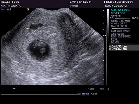
Another woman at 7 weeks with vaginal bleeding: Again, you see a subchorionic hematoma, but this time, it is just old blood outside the placenta. It does not harm the baby and usually gets reabsorbed as the pregnancy grows. In cases like these, hope is not lost—a repeat scan in two weeks often brings relief and reassurance.
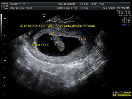
This is what every patient wants to see: A healthy 8-week pregnancy. The baby measures 20 mm (CRL), which is perfect for the gestational age. Seeing this image can finally let you breathe a little easier, knowing things are on track.
Why the Way the Scan Is Done Matters
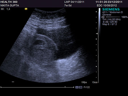
This is a 7-week healthy pregnancy, but the image was taken using a transabdominal scan (probe on the belly). Notice how the picture is not very clear. Now look at the next image:
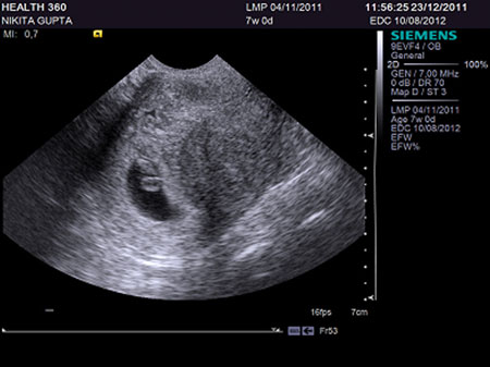
Here, the same pregnancy is scanned using a transvaginal probe. The difference is obvious: The image is much sharper. Transvaginal scans are now the standard for early pregnancy because they provide clearer, more reliable images. Rest assured, this method is safe for both you and your baby—the probe stays in the vagina, while your baby is safe in the uterus, far out of reach.
Early scans are best done transvaginally for accurate, detailed images—do not worry, it is safe for the baby.
Technology That Offers Reassurance
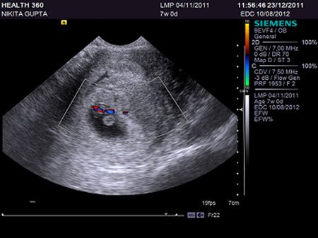
The use of color Doppler in ultrasound can show you blood flow within the pregnancy, proving the baby is getting nourishment and the placenta is working well.
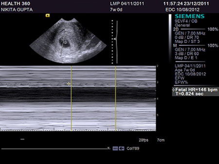
An M-mode Doppler can help your doctor show you your baby’s heartbeat. That flicker on the screen is often the first real proof of life—a moment many couples never forget. It is a strong sign that nature is doing what it should.
What Should You Do After the 2WW?
Here is the honest truth: After those first positive results, you will need a series of ultrasounds. And while each scan can bring hope, it can also raise new questions. Here is what you can do:
- If you see a gestational sac but no fetal pole before 6 weeks, do not panic. It is often just too early. Trust your doctor’s plan for repeat scans.
- Bleeding does not always mean disaster. Many women with subchorionic hematomas go on to have healthy pregnancies.
- If the scan is not clear, ask for a transvaginal scan.
- Remember: Growth from one scan to the next is what matters most. A single snapshot does not tell the whole story.
At Malpani Infertility Clinic, we believe in giving you all the facts and supporting you through the ups and downs. We guide you to understand your situation and help you make the best decisions for yourself. If you have any doubts, concerns, or simply want to talk through your scan results, do not hesitate to consult with Dr. Malpani for clear, compassionate advice—no sugar-coating, just honest guidance that puts you in control.
Frequently Asked Questions
Q: Why do I need multiple ultrasounds in early pregnancy?
A: Early pregnancy develops quickly, and a single scan often does not tell the full story. Serial ultrasounds help track your baby’s growth and pick up any issues as early as possible.
Q: My scan showed a gestational sac but no baby. Should I worry?
A: Not necessarily. Before 6 weeks, it is common to see only the sac. Your doctor will likely recommend another scan in a week to check for development.
Q: What does a subchorionic hematoma mean? Will I miscarry?
A: A subchorionic hematoma is a collection of blood in the uterus. Many women with these go on to have healthy pregnancies. The risk of miscarriage depends on size, location, and whether the baby is growing well.
Q: Is a transvaginal scan safe for my pregnancy?
A: Yes, it is completely safe. The probe does not reach the uterus, and it gives clearer images than a scan done on your belly.
Q: When should I be able to see my baby’s heartbeat?
A: The fetal heartbeat is usually visible by 6 to 7 weeks of pregnancy on a good-quality, transvaginal ultrasound.
Q: What if my baby measures behind the expected size?
A: Sometimes pregnancies start off slow but catch up. Your doctor will suggest follow-up scans to check for ongoing growth before making any decisions.
Q: What should I do if I am anxious about my scan results?
A: It is normal to feel anxious. If you need more support or a second opinion, reach out to an expert who will patiently explain your results and guide your next steps.