
Sonography Scans for Infertility Treatment. Ultrasound - Seeing with Sound
Have you ever sat in a waiting room, heart pounding, wondering if this next scan will finally give you some answers, hoping, dreading, and wishing you could understand what all those black and white shapes on the screen mean? If you are struggling to get pregnant or are feeling lost after repeated disappointments, you are not alone. So many couples find themselves confused and anxious about the tests and procedures, especially when it comes to ultrasound scans for infertility. Let’s cut through the uncertainty and talk honestly about what these scans can (and cannot) do for you, and why having the right clinic and doctor by your side can make all the difference.
How Ultrasound Brings Clarity to Infertility
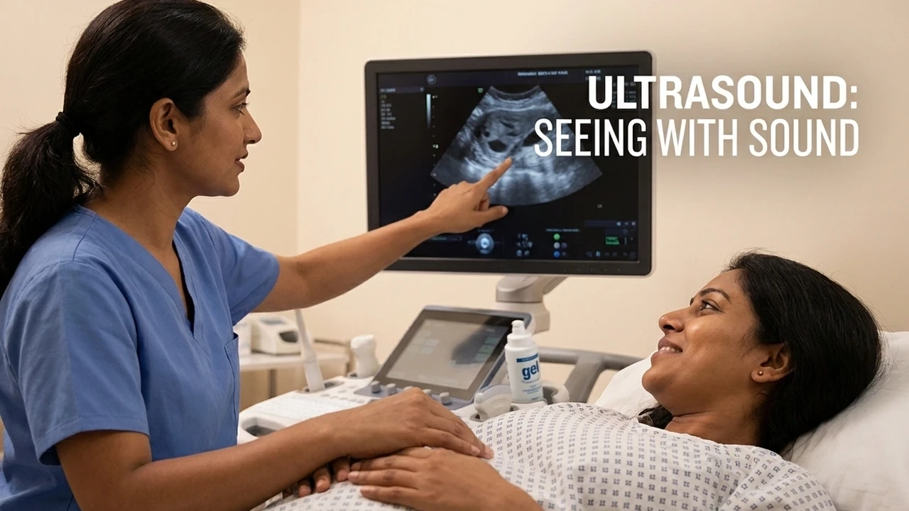
Ultrasound, sometimes called sonography, is not just another test. For couples facing infertility, it is a window into the hidden world inside the pelvis. Whether you are at the very beginning of your fertility journey or you have already tried multiple cycles, ultrasound scans help us see what is happening with the uterus and ovaries without any pain, surgery, or downtime. It is completely safe, using sound waves (not radiation), and allows us to literally “see with sound”.
Think of it like a highly sensitive, real-time camera that lets your doctor watch your body’s reproductive clockwork in action. The older abdominal ultrasound method required you to drink litres of water and hold your bladder until you thought you might burst. Thankfully, those days are mostly gone. Today, transvaginal (internal) ultrasound is the gold standard for infertility care. The slender probe is gently placed inside the vagina, bringing it much closer to the uterus and ovaries, so the images are clearer and more detailed. Most women find this much more comfortable, and far less awkward, than the old method.
What Ultrasound Really Shows, and What It Cannot
It is tempting to hope that a single scan will answer every question. The truth is, ultrasound is fantastic for some things, and limited for others. Here is what it does best:
- Visualizing the uterus: We can check for fibroids (muscular lumps that can block or distort the womb), polyps, or unusual shapes that might make pregnancy difficult.
- Assessing the ovaries: We can see the number and size of follicles (the tiny sacs that contain your eggs), look for cysts, and spot signs of conditions like polycystic ovary syndrome (PCOS) or endometriosis.
- Early pregnancy confirmation: The scan can pick up a pregnancy weeks before a urine test, and even spot problems like ectopic pregnancy early on.
- Endometrial lining: We check how thick and healthy the lining of your womb is, which matters for embryo implantation.
But there are limits. Ultrasound is not the best tool for seeing if your fallopian tubes are open and working. For that, specialized tests like an HSG (a dye test) or laparoscopy may be needed. Remember: A scan is only as good as the doctor interpreting it. Those blurry black and white images are not self-explanatory, they require real skill, experience, and context.
Follicular Scanning: Tracking Ovulation in Real Time
One of the most powerful uses of ultrasound is follicular monitoring, watching your ovaries as they prepare to release an egg. If you have ever wondered, “Am I ovulating? Is my egg growing at the right time? When is the best time for intercourse or for an IUI?” this is the test that provides real answers.
During a typical follicular study, you will have scans every few days (sometimes daily as ovulation approaches). The doctor will measure the size of your developing follicle. On the screen, it looks like a round, black bubble. When it reaches the right size (usually 18-22 mm), it is ready to ovulate. Sometimes, you can actually watch the follicle rupture and release the egg, an incredible moment of clarity in a journey full of question marks.
But that is not all. These scans also let us measure your endometrial lining, the “bed” where an embryo needs to implant. A healthy lining is thick, plush, and shows a distinct triple-layered appearance. If it is too thin, too bright, or irregular, it might signal a hormonal problem or a barrier to pregnancy.
 Fig 1. Ultrasound scan showing multiple follicles
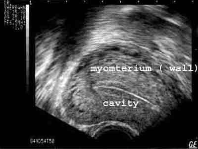 Fig 2. Ultrasound scan of the uterus, showing a normal endometrium, which appears as a triple band in the center of the uterus
Seeing your own follicles and lining develop on the screen can transform anxiety into understanding, you know exactly what is happening, and when.
Ovarian Cysts: Should You Worry?
Few things are more stressful than hearing the word “cyst” during a scan. But here’s the honest truth: most ovarian cysts found during infertility workups are harmless and temporary. They are simply fluid-filled sacs, often related to the normal process of ovulation. For example, sometimes a follicle grows but does not release the egg, this is called a follicular cyst. Or after ovulation, the space where the egg was released fills with blood and forms a corpus luteum cyst. Both are considered functional cysts and typically shrink away on their own within a few weeks.
Occasionally, more complex cysts are seen. These might have thicker walls, internal echoes, or look “messy” on the scan. Examples include endometriotic cysts (sometimes called chocolate cysts) or, rarely, ovarian tumors. These need closer monitoring or, sometimes, further testing.
- Functional cysts usually disappear within 1-2 menstrual cycles and rarely need treatment.
- If a cyst is larger than 6 cm, or sticks around for more than 6 weeks, your doctor may recommend further evaluation.
- Certain fertility medications (like clomiphene or HMG) can temporarily increase the risk of cysts.
What matters most is that your doctor knows how to interpret the scan in the context of your symptoms, cycle day, and treatments. At Malpani Infertility Clinic, we believe in empowering you with clear information, so you never feel left in the dark.
Who Should Perform Your Infertility Ultrasound?
This is a question many patients never think to ask, but it can make a huge difference to your care. While any radiologist or sonographer can technically perform a scan, not everyone can interpret the images in the context of your unique fertility story. The best results come when your infertility specialist personally performs or reviews your scans. Why? Because treatment decisions, when to trigger ovulation, whether to delay a cycle, how to adjust your medications, depend on the nuances seen on those images in real time.
At Malpani Infertility Clinic, we insist on performing and reviewing your scans ourselves. This means:
- You get immediate feedback and advice, not a report you need to wait for.
- We can adapt your treatment protocol instantly, without delays or confusion.
- If we spot anything unusual, we can explain it right away, often showing you the images and answering your questions on the spot.
And if you live far away, we can even interpret emailed images securely, thanks to advances in telemedicine. You should never have to run from the sonographer to the gynecologist, only to feel more confused and anxious.
Frequently Asked Questions
Q: Is an ultrasound scan painful or risky?
A: Transvaginal ultrasound is generally not painful, though you may feel mild pressure. It is completely safe and does not use radiation. Most patients find it far more comfortable than older abdominal scans that required a full bladder.
Q: How often will I need ultrasound scans during infertility treatment?
A: The number of scans depends on your treatment plan. For ovulation monitoring, you may need several scans in one cycle. For IVF or IUI cycles, even more frequent scans might be necessary to track follicle growth and lining development.
Q: What if a cyst is found on my ultrasound?
A: Most cysts are functional and disappear within 1-2 cycles. Your doctor will monitor the cyst and only intervene if it grows large, persists, or looks suspicious. You will be kept fully informed at each step.
Q: Can ultrasound scans detect blocked fallopian tubes?
A: Ultrasound cannot reliably show if tubes are blocked. For this, special tests like HSG or laparoscopy are required. However, ultrasound can sometimes spot indirect signs, like fluid around the tubes.
Q: Who is the best person to perform my infertility ultrasound?
A: Your scans should ideally be performed and interpreted by a fertility specialist who understands your personal history and treatment plan, not just a general radiologist. This ensures the results are meaningful and actionable for your care.